I. 3D-C-assisted navigation surgery-FESS-of the nose, paranasal sinuses and the scull base, with the use of computer assisted (CA)-RP (rapid prototyping) models (3D-CA-RP-FESS)
Dg. Rhinosinuitis chronica polyposa (polypus antrochoanalis sinus paranasales lateris dextri/sinus frontalis, ant/post ethmoidalis, maxillaris). Fractura multipl. septi na¸si cum deviationem palatinalis septi nasi lateris dextri (lamina qvadrangularis et perpendicularis) ar III-V. Perforatio septi nasi ar III. Hypertrophia conharum nasalis inferioris nasi bilateralis.
Op.: Endo-op. (cum reconstructionem) functionalis septi nasi, cum plasticam perforationis septi ar III. Operatio sinus paranasales per viam 3D-CAS-endoscopicam (3D-CA-FESS).
Op.: Endo-op. (cum reconstructionem) functionalis septi nasi, cum plasticam perforationis septi ar III. Operatio sinus paranasales per viam 3D-CAS-endoscopicam (3D-CA-FESS).
Interactive display of correlated 2D and 3D data in a four-window format may assist the endoscopist in performing various image guided procedures. In comparison with real endoscopy, the VE is completely non-invasive. It is possible to repeat the same procedure several times, therefore it may be a valuable tool for training, as well as the interactive control of all virtual camera parameters, including the field-of-view, and the viewing as opposed to the extend of lesions within and beyond the wall which gives the potential to stage tumors by determining the location and the extent of transmural extension. In the nasal or sinus cavity, VE can clearly display the anatomic structure of the paranasal sinuses, nasopharyngeal cavity and upper respiratory tract, revealing damage to the sinus wall caused by a bone tumor or fracture, and use the corresponding cross-sectional image or multiplanar reconstructions to evaluate structures outside the sinus cavity. A major disadvantage of VE is its inability to make an impact on operating room performance, as well as the considerable time consumption, to evaluate the mucosal surface, or to provide a realistic illustration of the various pathologic findings in cases with highly obstructive sinonasal disease. Considering the specificities and basic features of 3D-CA-navigation surgery and tele-3D-CAS, we believe that this type of surgery would be acceptable to many surgeons all over the world .The possibility of data analysis and storage in the 3D form and development of 3D centers at clinical institutions, as well as the development of the surgery using a remote-controlled robots should provide a new quality in proper training of future surgeons in 3D-CAS as well as tele-3D-CAS activities. Finally, modelling of the biological material and tissue properties is an important field of research. In the future, we can expect a new generation of diagnostic imaging techniques that use simulated reality techniques for effective visualization of organ anatomy and function. Such systems will enable not only better medical diagnosis but also more appropriate intervention.

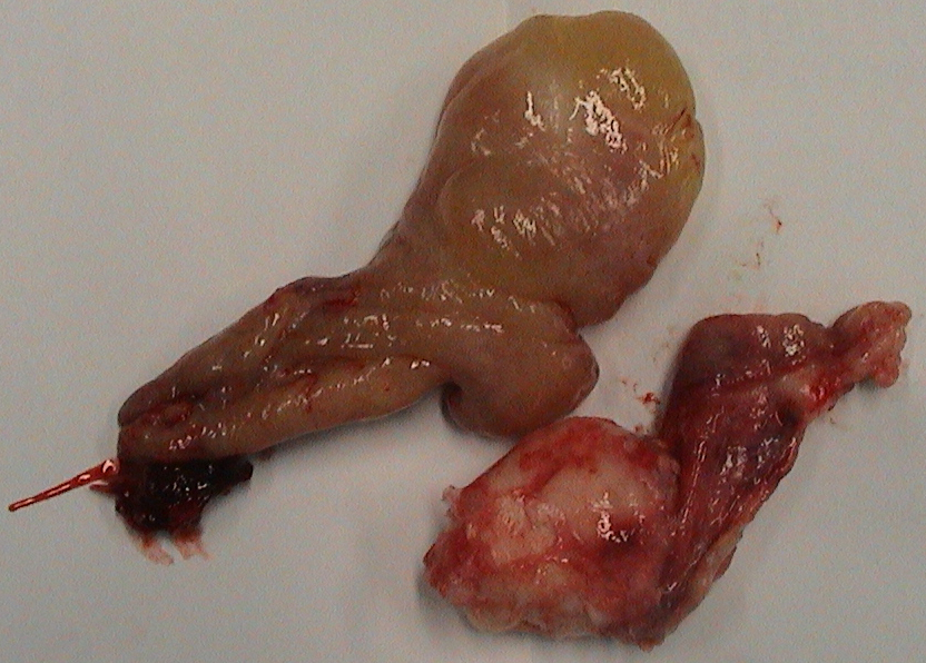


Figure 1. Visualization of patho-tissue in comparison with the head/nose of the patient (Figures 1.a,b), and 3D models (RP models) of the human head in different projections (Figures 1.c,d). Visualization of all paranasal sinuses and surrounding regions from different 3D aspects. Virtual 3D models also can be used in actual reality for tangible, physical (real) models obtained e.g.by rapid prototyping (additive manufacturing technique). Rapid Prototyping (RP) techniques look most promising to satisfy medical need for tangible models. While prototyping is a usually slow and expensive process of building pre-production models of a product to test various aspects of its design, rapid prototyping techniques are methods that allow quick production of physical prototypes.
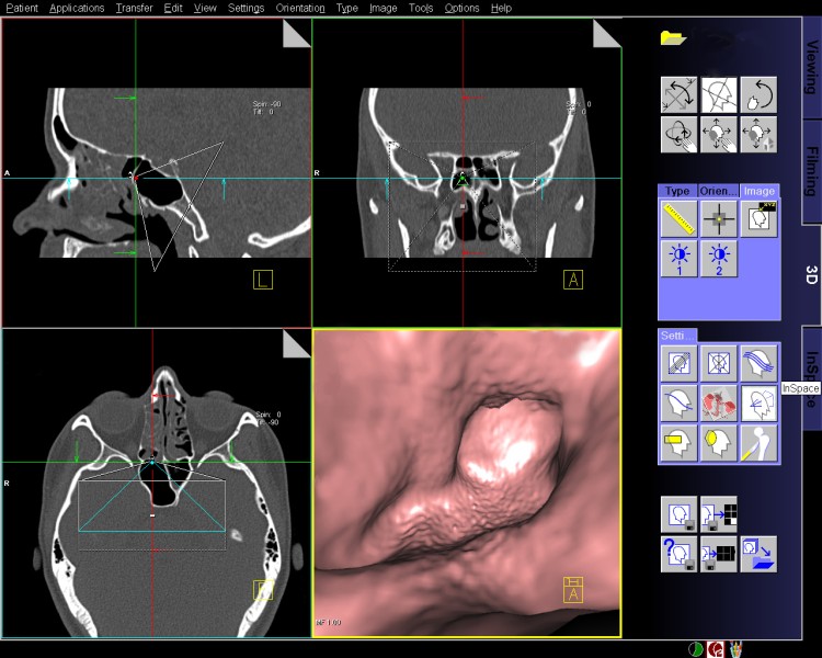
Figure 2. We performed VE of paranasal sinuses, and nasal cavity, as a part of diagnostic or preoperative management.