23rd Congress of the European Rhinologic Society, and the 29th International Symposium on Infection and Allergy of the Nose, Geneva, Switzerland, 23rd ERS & 29th ISIAN, June 2010
Symposium
Robotics and new technology
Image guided surgery (neuronaviation)
Intraoperative and/or in-office CT imaging
Laser in rhinology
Shaver in FESS
Invited speakers: M.D. Caversaccio (CH), D. Kennedy (USA), I. Klapan (CRO),
L.P. Nolte (CH), G. Strauss (D), P. Hermann (D)
Wednesday
23 June, 2010
8.00-10.00 I. Klapan (Zagreb, CROATIA), Faculty, Invited speaker
Application of advanced virtual reality (VR) and 3D computer
assisted technologies in rhinology: CAS and Tele-CAS
Objectives: 3D image analysis and processing, tissue modelling, virtual endoscopy and surgery (VE, VS), 3D-computer assisted surgery (3D-CAS), as well as tele-3D-CAS, represent a basis for various realistic simulations in medicine, and can definitely create an impression of immersion of a physician in a non-existing virtual environment. In rhinology, such an revolutionary approach is of paramount importance for the surgeon because of the proximity of intracranial structures and limited operative field layout hampering spatial orientation during the “standard” operative procedure. The possibility of exact preoperative, non-invasive visualization of the spatial relationships of anatomic and pathologic structures, including extremely fragile ones, size and extent of pathologic process, and of precisely predicting the course of surgical procedure, allows the surgeon to achieve considerable advantage in the preoperative examination of the patient and to reduce the risk of intraoperative complications, all this by use different VR methods. Methods: From the very beginning of our 3D-CAS and tele-3D-CA-surgeries, the modeling was done by use of the VolVis, Volpack/Vprender, GL Ware programs on a DEC Station 3100 computer. With the advent of 3D Viewnix V1.0 software, we started using this program, and then 3D Viewnix V1.1 system, AnalyzeAVW system, T-Vox system and OmniPro 2 system on Silicon Graphics O2, Origin200 and Origin2000 computers. Our team used several standards to encode live video signals in telesurgery, such as M-JPEG, MPEG1, MPEG2 and MPEG4. For conferencing/consultation cameras used between two or more connected sites during the surgery, we used JPEG and MPEG1 stream with audio. ORs were connected using several computer network technologies with different bandwidths, from T1, E1 and multiple E1 to ATM-OC3 (from 1Mb/s to 155Mb/s). For computer communications using X-protocol for image/3D-models manipulations, we needed an additional 4Mb/s of bandwidth, instead of the 1Mb/s when we used our own communication tools for the transfer of surgical instrument movements. Discussion: The real-time requirement means that the simulation must be able to follow the actions of the user that may be moving in the virtual environment. The computer system must also store in its memory a 3D model of the virtual environment (3D-CAS models). In that case a real-time virtual reality (VR) system will update the 3D graphical visualization as the user moves, so that up-to-date visualization is always shown on the computer screen. Upon the completion of the CAS and/or tele-CAS-operation, the surgeon compares the preoperative and postoperative images and models of the operative field, and studies video records of the procedure itself. In otorhinolaryngology, especially in rhinology, research in the area of 2D and 3D image analysis, visualization, tissue modelling, and human-machine interfaces provides scientific expertise necessary for developing successful VR applications. The basic requirement in rhinology, resulting from the above mentioned needs refers to the use of a computer system for visualization of anatomic 3D-structures and integral operative field to be operated on. To understand the idea of 3D-CAS/VR it is necessary to recognize that the perception of surrounding world created in our brain is based on information coming from the human senses and with the help of a knowledge that is stored in our brain. The usual definition says that the impression of being present in a virtual environment, such as virtual endoscopy (VE) of the patient’s head, that does not exist in reality, is called VR. The user/physician, has impression of presence in the virtual world and can navigate through it and manipulate virtual objects. A 3D-CAS/VR system may be designed in such a way that the physician, is completely immersed in the virtual environment. Conclusions: VR applications as well as 3D reconstruction of anatomic units becomes a routine preoperative procedure, as we already shown in our surgical activities in the last two decades (our first CAS/May 1994, tele-3D-CAS/October 1998), providing a highly useful and informative visualization of the regions of interest, thus bringing advancement in defining the geometric information on anatomical contours of 3D-human head-models by the transfer of so-called “image pixels” to “contour pixels”. The possibility of data analysis and storage in the 3D form and development of 3D centers at clinical institutions should provide a new quality in proper training of future surgeons in CAS as well as tele-CAS activities (www.mef.hr/MODERNRHINOLOGYand www.poliklinika-klapan.com).
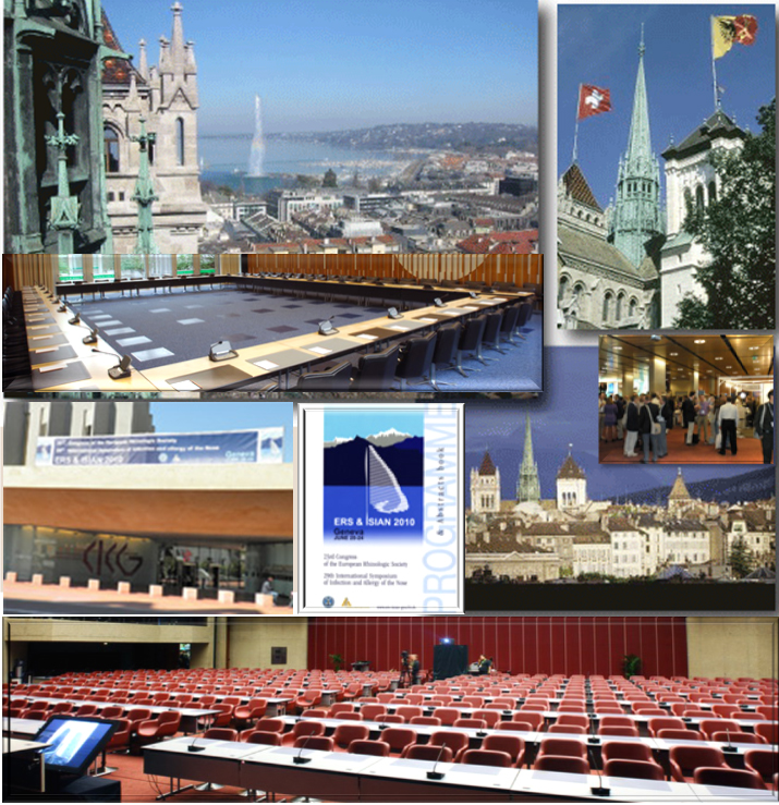
23rd ERS & 29th ISIAN, Geneva, Switzerland 2010.
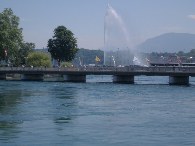
A) 23rd ERS&ISIAN Congress Center, and B) the City of Geneva
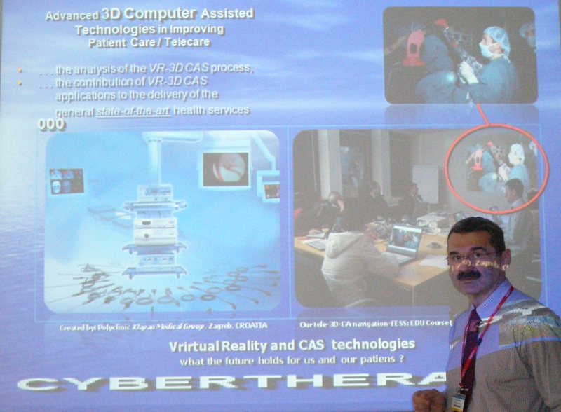
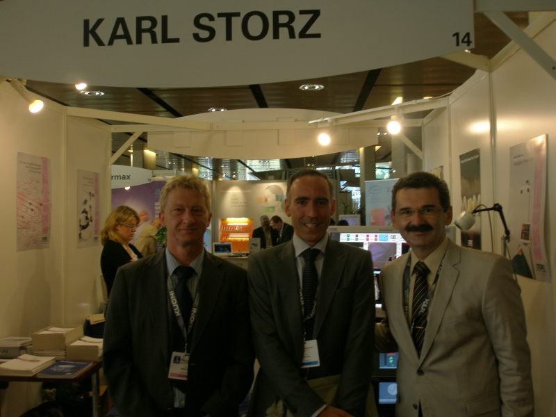
Geneva, Switzerland, 23rd ERS & ISIAN. A) Professor Klapan (Instructional Course/faculty/invited speaker), and B) with representative of Cart Storz Co., and Professor G. Strauss, Germany