24th Congress of the European Rhinologic Society, and the 31st International Symposium on Infection and Allergy of the Nose, Toulouse, FRANCE, 24th ERS & 31st ISIAN, June 2012
|
Monday, 18 June
2012. |
Virtual reality and computer integrated surgery in rhinology
I. Klapan (Zagreb, CROATIA), Chairman, Instructional Course
|
|
|
|
||
Virtual Reality (VR) and computer integrated surgery in rhinology
Modern medical 3D-image processing software (SW) covers most of medical imaging methods like MRI, MSCT, PET and microscopy, scientific and other industrial imaging applications. 3D-surface models or volume rendering from 2D-cross-section images once created in real time with SW can be used for measuring, operation planning, simulation, structural finite element analysis, uas well as for various Virtual Reality applications. Nowadays, 3D image processing SW is successfully used in many medical cases in navigation surgery, 3D-CA-surgery, tele-3D-CA-surgery as well as in virtual endoscopy (VE) and virtual surgery (VS). Furthermore, the 3D models also can be used for physical medical models obtained e.g. by rapid prototyping (additive manufacturing) technique. Rapid Prototyping (RP) techniques look most promising to satisfy medical need for tangible models. While prototyping is a usually slow and expensive process of building pre-production models of a product to test various aspects of its design, rapid prototyping techniques are methods that allow quick production of physical models. In this Instructional Course, we are introducing guidelines and suggestions for use of 3D image processing SW in head and neck pathology diagnostic and procedures for obtaining physical medical model by additive manufacturing/rapid prototyping techniques, bearing in mind the improvement of surgery performance, its maximum security and faster postoperative recovery of patients. Different VR applications become a routine preoperative procedure in human medicine, as we already shown in our surgical activities in the last two decades (from June 03, 1994), providing a highly useful and informative visualization of the regions of interest, thus bringing advancement in defining the geometric information on anatomical contours of 3D-human head-models by the transfer of so-called “image pixels” to “contour pixels”.
Telemedicine attempts to break the distance barrier between the provider and the patient in health-care delivery. VR is able to simulate remote environments and can therefore be applied to telemedicine. Physicians can have VR produced copy of a remote environment including the patient at their physical location. One of the simplest telemedical applications is medical teleconsultation, where physicians exchange medical information, over computer networks, with other physicians in the form of image, video, audio, and text. Teleconsultations can be used in radiology, pathology, surgery, and other medical areas. One of the most interesting telemedical applications is tele-surgery. Telesurgery is a telepresence application in medicine where the surgeon and the patient are at different locations, but such systems are still in an early research phase. Patients, who are too ill or injured to be transported to a hospital, may be operated remotely. In all these cases, there is a need for a surgeon specialist who is located at some distance. Generaly speaking, the purpose of a tele-presence system is to create a sense of physical presence at a remote location. Tele-presence is achieved by generating sensory stimulus so that the operator has an illusion of being present at a location distant from the location of physical presence. A tele-presence system extends operator’s sensory-motor facilities and problem solving abilities to a remote environment. A tele-operation system enables operation at a distant remote site by providing the local operator with necessary sensory information to simulate operator’s presence at the remote location. Tele-operation is a special case of tele-presence where in addition to illusion of presence at a remote location operator also has the ability to perform certain actions or manipulations at the remote site. In this way it is possible to perform various actions in distance locations, where it is not possible to go due to a danger, prohibitive price, or a large distance.Realization of VR systems require software (design of VE) for running VR applications in real-time. Simulations in real-time require powerful computers that can perform real-time computations required for generation of visual displays.
Different goals can be achieved by using different VR applications. These goals range from teaching, diagnosis, intervention planning, providing insight into the potentially complicated and non-standard anatomy, as well pathology of the patients, intra-operative navigation, etc. VE or fly-through methods which combine the features of endoscopic viewing and cross-sectional volumetric imaging provided more effective and safer endoscopic procedures in diagnostics and management of our patients, especially preoperatively, as we already discussed. This approach can also be applied for training and familiarize the surgeon with endoscopic anatomic appearance. Definitely, the presentation of image data in such a way enables the operator not only to explore the inner wall surfaces but also to navigate inside the virtual organs extracted from MSCT and/or MR images of nasal cavity, paranasal sinuses and scull base, and in combination with in-space skull bone rendering, offers plastic and accurate additional 3D information for head and neck surgeon in combination with classical 2D black and white CT images. Interactive display of correlated 2D and 3D data in a four-window format may assist the endoscopist in performing various image guided procedures. In comparison with real endoscopy, the VE is completely non-invasive. It is possible to repeat the same procedure several times, therefore it may be a valuable tool for training, as well as the interactive control of all virtual camera parameters, including the field-of-view, and the viewing as opposed to the extend of lesions within and beyond the wall which gives the potential to stage tumors by determining the location and the extent of transmural extension. In the nasal or sinus cavity, VE can clearly display the anatomic structure of the paranasal sinuses, nasopharyngeal cavity and upper respiratory tract, revealing damage to the sinus wall caused by a bone tumor or fracture, and use the corresponding cross-sectional image or multiplanar reconstructions to evaluate structures outside the sinus cavity. A major disadvantage of VE is its inability to make an impact on operating room performance, as well as the considerable time consumption, to evaluate the mucosal surface, or to provide a realistic illustration of the various pathologic findings in cases with highly obstructive sinonasal disease.
Even more, our vision of 3D-CA-navigation surgery and/or tele-3D-CA-navigation surgery allows surgeons not only to see and transfer video signals but also to transfer 3D computer models and surgical instrument movements with image/3D-model manipulations in real time during the surgery. Considering the specificities and basic features of 3D-CA-navigation surgery and tele-3D-CAS, we believe that this type of surgery would be acceptable to many surgeons all over the world for the following reasons: a) the technology is readily available in collaboration with any telecom worldwide, b) the improved safety and reduced cost will allow the inclusion of a greater number of patients from distant hospital institutions in such a telesurgical expert system, c) the “presence” of leading international surgical experts as tele-consultants in any OR in the world will thus be possible in the near future, which will additionally stimulate the development of surgery in all settings; and d) the results obtained in the tele-3D-CAS project in Croatia are encouraging and favor the further development of the method. The possibility of data analysis and storage in the 3D form and development of 3D centers at clinical institutions, as well as the development of the surgery using a remote-controlled robots should provide a new quality in proper training of future surgeons in 3D-CAS as well as tele-3D-CAS activities (www.mef.hr/MODERNRHINOLOGY andwww.poliklinika-klapan.com). Finally, modelling of the biological material and tissue properties is an important field of research. In the future, we can expect a new generation of diagnostic imaging techniques that use simulated reality techniques for effective visualization of organ anatomy and function. Such systems will enable not only better medical diagnosis but also more appropriate intervention.
Keywords: Virtual reality, Virtual endoscopy, Additive manufacturing, Rapid prototyping, Medical models, Medical imaging, Endoscopy, 3D imaging, Rhinology, Paranasal sinuses, Health, Engineering


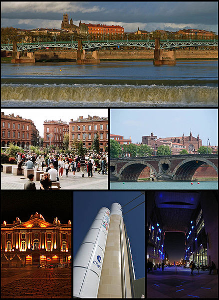
Toulouse, France, 24th ERS & 31st ISIAN, June 2012.
A/B) Professor Klapan, Instructional Course RHINOLOGY/faculty/invited speaker


Toulouse, France, 24th ERS & 31st ISIAN, June 2012. A) Congress Center, B) The City of Toulouse